PrepSuite.co.uk


Try 5 Free NEBDN Radiography Questions
Get a feel for the questions by taking a quick practice test below.
Question 1

A clinician reviews the radiograph provided to assess periodontal bone levels. What is the most appropriate radiographic type for this assessment?
A. Periapical radiograph using paralleling technique
B. Horizontal bitewing radiograph
C. Occlusal radiograph
D. Panoramic radiograph
E. Vertical bitewing radiograph
Answer: E Vertical bitewings are preferred for assessing periodontal bone levels as they provide greater vertical coverage of alveolar bone compared to horizontal bitewings.
Question 2
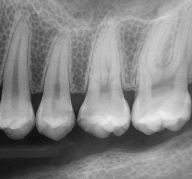
A student incorrectly identifies this radiograph as a bitewing. What is the most significant error in their interpretation?
A. Failure to recognise the presence of restorations
B. Incorrect assumption that only crowns are visible
C. Misinterpretation of trabecular bone pattern
D. Confusion between anterior and posterior teeth
E. Overestimation of radiographic contrast
Answer: B The image clearly shows full roots and apices, not just crowns, which distinguishes it from a bitewing radiograph.
Question 3
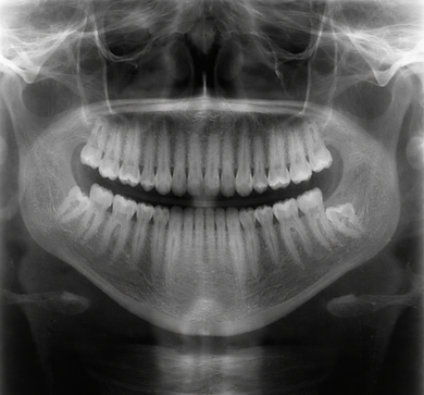
A patient undergoes this radiographic examination. Which sequence best describes the process of acquiring this image?
A. Place sensor intraorally → align X-ray beam → expose image
B. Position patient → rotate X-ray source and detector around the head → capture composite image
C. Insert film parallel to tooth → direct beam perpendicular → expose image
D. Place film on occlusal surface → direct beam vertically → expose image
E. Position receptor behind tooth → bisect angle → expose image
Answer: B Panoramic radiography involves positioning the patient while the X-ray source and detector rotate around the head to produce a composite image of the jaws.
Question 4
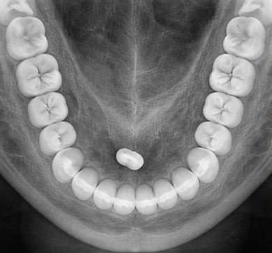
A student incorrectly identifies this radiograph as a panoramic image. What is the key feature that contradicts this interpretation?
A. Presence of both maxillary and mandibular teeth
B. Limited view confined to the mandibular arch
C. High level of distortion across the image
D. Absence of root structures
E. Uniform density of bone throughout
Answer: B Panoramic radiography involves positioning the patient while the X-ray source and detector rotate around the head to produce a composite image of the jaws.
Question 5
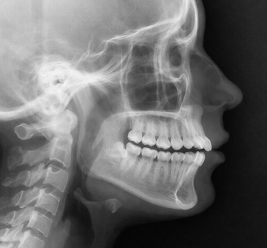
A clinician requests this radiograph to assess skeletal relationships prior to orthodontic treatment. Why is this radiographic type most appropriate?
A. It provides detailed inter proximal caries detection
B. It allows evaluation of soft tissue profile and jaw relationships
C. It shows high-resolution images of individual root apices
D. It captures both arches simultaneously in a curved plane
E. It minimises distortion in posterior teeth only
Answer: B Lateral cephalometric radiographs are used to assess skeletal and soft tissue relationships, making them essential in orthodontic planning.